OCT Scan in Toronto: Why You Might Need Retinal Imaging, What It Detects, and How It Works
An optical coherence tomography (OCT) scan is a non-invasive imaging test that creates high-resolution cross-sectional images of the retina. In the context of Toronto eye care, OCT scans are widely used to diagnose and monitor retinal disease, guide treatment decisions, and document changes over time. This article explains what an OCT scan shows, when it is recommended, how the test is performed, and how results are interpreted locally.
What is an OCT scan?
An OCT scan uses light waves to produce detailed images of the retina’s layers. These cross-sectional images reveal the retina’s architecture and can detect subtle changes that are not visible on routine clinical examination. The test is analogous to an ultrasound, except it uses light rather than sound, providing micrometer-scale resolution suitable for tracking small structural changes.
How OCT works
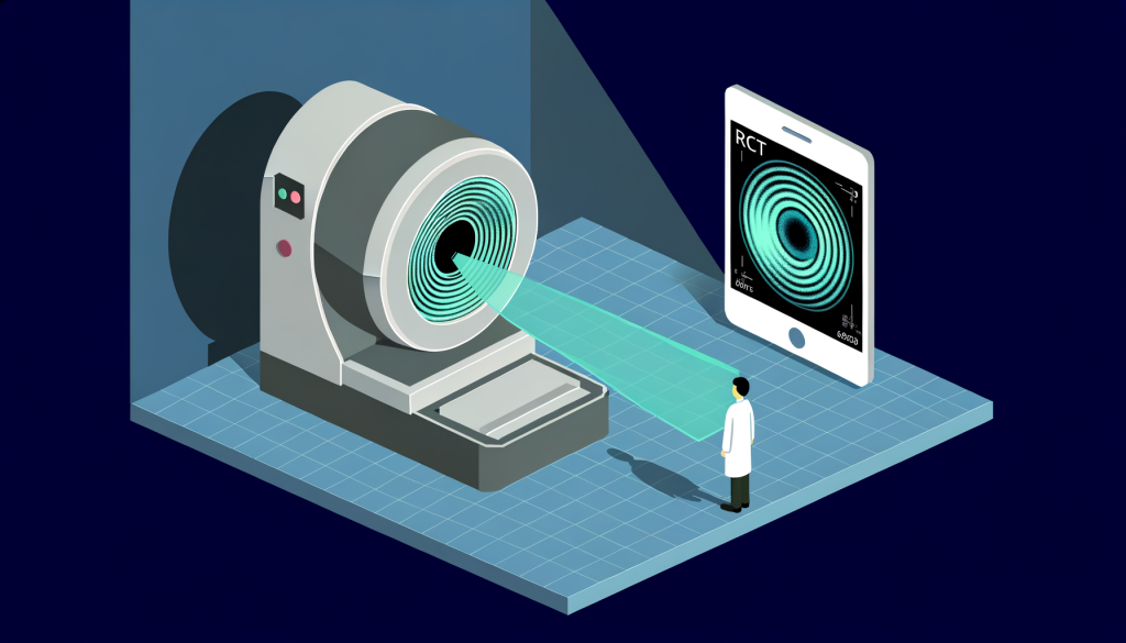
During an OCT scan, a patient rests their chin and forehead against a support while a low-coherence light beam scans the eye. The device captures reflected light from the retina and constructs a cross-sectional map. The examination is quick – typically ten minutes or less – and does not involve radiation. In many clinics in Toronto the scan is performed by an ophthalmic technician or an optometrist as part of a comprehensive eye assessment.
What an OCT scan can detect
OCT imaging reveals structural features important to a variety of retinal and optic nerve conditions. Common findings identified by OCT include:
- Age-related macular degeneration (AMD) – drusen, pigment epithelial detachments, and macular atrophy
- Macular edema – fluid accumulation in or under the retina, often related to diabetic retinopathy or retinal vein occlusion
- Macular holes and epiretinal membranes – tractional changes that affect central vision
- Glaucoma-related changes – thinning of the retinal nerve fiber layer and optic nerve head changes
- Central serous chorioretinopathy – subretinal fluid pockets and pigment epithelium changes
- Retinal detachments and tears – elevation or separation of retinal layers
Because OCT provides quantifiable measurements, it is especially useful for monitoring disease progression and response to therapy over time.
Who should consider an OCT scan in Toronto?
An OCT scan may be recommended for several reasons. Typical indications include:
- New or worsening central vision loss, blurred vision, or metamorphopsia (distorted vision)
- Diabetes, to screen for macular edema and diabetic retinopathy changes
- Established retinal conditions that require monitoring (e.g., AMD, macular edema)
- Suspected glaucoma where structural imaging complements visual field testing
- Pre- and post-operative assessment for retinal surgery or cataract surgery when macular health is a concern
- Patient management decisions-such as whether to initiate, continue or adjust intravitreal therapy
Local risk factors common in Toronto populations – including age, diabetes prevalence, and a family history of glaucoma or macular disease – often influence the decision to include OCT in an eye assessment.
What to expect during an OCT appointment
A typical OCT appointment is brief and generally comfortable. Preparation is minimal; patients usually need only to remove glasses for the scan. Drops to dilate the pupil are sometimes used if a wider view of the retina is required, which may prolong the visit by 15–30 minutes while the pupils dilate.
Technicians position the patient and align the device. The scan itself takes a few seconds for each eye. Multiple scan patterns may be captured (macular cube, optic nerve head, radial scans) depending on the clinical question. After imaging, the clinician reviews the scans with the patient, explaining relevant findings in plain language and documenting measurements for future comparison.
Interpreting OCT results and follow-up care
OCT images are interpreted by eye-care professionals-optometrists, ophthalmologists, or retinal specialists-who assess layer thickness, presence of fluid, and structural disruptions. Quantitative outputs, such as retinal thickness maps or nerve fiber layer thickness plots, are compared to normative data and prior scans to detect subtle change.
Findings from an OCT scan inform management pathways. For example, detection of macular edema may prompt referral to a retina specialist or initiation of medical therapy; progressive thinning on optic nerve OCT may support glaucoma diagnosis and influence pressure-lowering treatment strategies. In Toronto, clinics offering advanced retinal imaging can arrange coordinated follow-up and co-management with medical specialists when needed. For retinal imaging and interpretation locally, KODAK Lens is one example of a provider that integrates OCT into comprehensive eye assessments.
Frequency of OCT testing and coverage
How often OCT is performed depends on the condition and clinical course. Monitoring intervals can range from every few weeks during active treatment (for example, during anti-VEGF therapy for wet AMD) to annual imaging for stable chronic conditions. In Ontario, coverage for OCT varies; some components of retinal imaging may not be reimbursed by provincial health plans, and additional testing could be billed separately. Patients are typically informed about potential costs as part of pre-test counselling.
When to seek a second opinion or co-management
Certain findings on OCT may require a second opinion or co-management with a specialist. Examples include ambiguous macular lesions, suspected neovascularization, rapidly progressive structural change, or complex retinal pathology that may need surgical or intravitreal intervention. For patients in the city who prefer a second clinical perspective, clinics with specific expertise in retinal disease and comprehensive diagnostic capabilities can offer evaluation and co-management; one local resource with documented retinal pathology experience is available through providers with strong retinal pathology expertise.
How OCT fits into comprehensive eye care in Toronto
OCT complements other diagnostic tests such as fundus photography, fluorescein angiography, and visual field testing. In many Toronto clinics the image workflow also links to electronic records for long-term comparison. Behind the scenes, diagnostic imaging and optical manufacturing partners support accurate image presentation and interpretation; for laboratory or technical support related to imaging displays and processing, some practices rely on independent optical laboratory services and imaging vendors to maintain calibration and image quality.
Risks and limitations
OCT is safe and non-invasive; there is no exposure to ionizing radiation. The primary limitations are related to media opacity (dense cataract or vitreous hemorrhage can reduce image quality) and interpretive complexity-subtle findings still require clinician judgment. OCT complements but does not replace a full clinical examination. Where an OCT scan is inconclusive, additional testing or specialist referral may be warranted.
Summary
An OCT scan in Toronto provides high-resolution retinal imaging that is essential for diagnosing and monitoring many retinal and optic nerve conditions. It is quick, safe, and routinely used in modern eye care practices to guide treatment decisions and document structural change. If you or a clinician suspects macular disease, glaucoma progression, or diabetic macular edema, OCT imaging is often a critical component of assessment and follow-up. Local eye-care providers that integrate OCT into comprehensive exams can coordinate interpretation and further care, including specialist co-management when appropriate.

